Understanding Breast Density
Did your mammogram results mention that you have dense breast tissue? In January 2015, Missouri passed a law requiring Mammography departments to inform patients on their breast density.
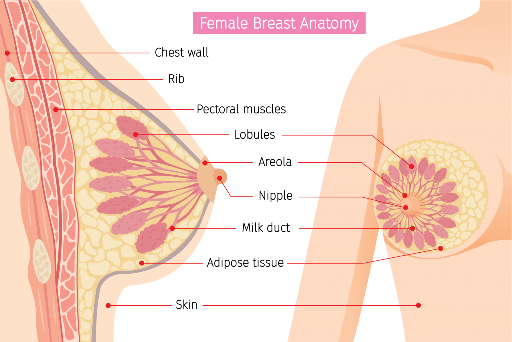
What is your breast anatomy?
Breasts are made up of lobules, ducts, fatty and fibrous connective tissue.
Lobules produce milk and are often called glandular tissue.
Ducts are tiny tubes that carry milk from the lobules to the nipple.
Fibrous and fatty (adipose) tissue give breasts their size and shape and hold the other tissues in place, your breasts will be seen as dense if you have a lot of fibrous or glandular tissue and not much fat in the breasts.
What is Breast Density?
Breast density compares the amount of fat to the amount of tissue on a mammogram. Dense breasts contain more glandular and fibrous tissue than fatty tissue. In general, younger women tend to have dense breasts, and breast density typically decreases as a woman gets older.
Women who have dense breasts are four to five times more likely to develop breast cancer than women with low breast density. Researchers are still trying to figure out why. We know that dense breasts can make it more difficult to find breast cancer on a mammogram. Since both cancer and dense breast tissue look white or light gray on a mammogram, the dense tissue may hide a tumor from view.
The Four Density Categories.

This chart shows us how radiologists “score” the tissue seen on a mammogram:
A-Almost Entirely Fatty: Breasts are almost all fatty tissue, easier to detect breast cancer.
B-Scattered Fibroglandular: There are scattered areas of dense glandular and fibrous tissue.
C-Heterogeneously Dense: More of the breast is made of dense glandular and fibrous tissue. This can make it hard to see small tumors in or around the dense tissue.
D-Extremely Dense: Breasts are extremely dense, which makes it hard to see tumors in the tissue.
Why does breast density matter in regards to a mammogram?
Dense breast tissue on a mammogram looks white, and breast masses or tumors also appear white on a mammogram; therefore, making it more difficult to detect breast cancer early. Breast density is very common. Some women have denser breast tissue than others. For most women, breasts become less dense with age, but in some women, there’s little change.
Why 3D mammography?
At Western Missouri Medical Center (WMMC), we suggest that every woman at the recommended screening age should consider 3D mammography instead of a regular 2D mammogram. A 3D mammogram provides more information of a woman’s breast tissue by examining the tissue one thin layer at a time by performing a sweep over the breast while under compression and can detect breast cancer at an earlier stage – saving more lives.
What is breast ultrasound?
Breast ultrasound is non-invasive and often used as a follow-up test after an abnormal finding on a mammogram, using sound waves, ultrasound is able to better categorize a mass seen on a mammogram.
Studies show breast ultrasound alone is not a good breast cancer screening tool. Therefore, ultrasound imaging can help to determine if an abnormality is solid (which may be a non-cancerous lump of tissue or a cancerous tumor) or fluid-filled (such as a benign cyst) or both cystic and solid. The only time an ultrasound should be performed before or instead of a mammogram is when a patient is under age 35 and/or pregnant and/or breastfeeding as ultrasound uses no radiation. Ultrasound used with mammography increases the detection of small tumors in patients with dense breasts. However, breast ultrasound does not replace mammography. With that being said, mammography overlooks roughly 10% of cancers in women with dense breast tissue.
What is a whole-breast ultrasound?
Whole-breast ultrasound (an ultrasound of the entire breast) is a technology that can be used with mammography to screen the breast for breast cancers that cannot be felt on physical examination or cannot be seen with mammography. The whole-breast ultrasound is designed to improve the detection rate of breast cancer in women with dense breasts, beyond what a mammogram alone can find. Whole-breast ultrasound should be done in addition to, but not replace, the screening mammogram.
WMMC provides this technology as a screening tool for women with dense breast tissue seen on a mammogram and for women under the age of 35 with a strong family history of breast cancer in a relative within 10 years of the patient’s age. We also provide this as a diagnostic tool for women feeling a lump if under 35 and/or pregnant and/or lactating.
How can I schedule my screening mammogram at WMMC?
If you are considering annual mammogram screenings, WMMC is here for you. To schedule an appointment, call (660) 262-7370 Monday – Friday 8:00 am – 5:00 pm.
Please be prepared to validate your personal and insurance information at the time of the call.
WMMC is proud to be a leading diagnostic facility in the West Central Missouri area. We hope that you will choose us as your partner for breast health, closer to home.